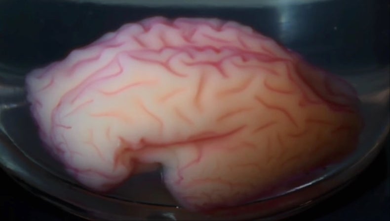
The human brain, a jelly-like mass of about 3 pounds, is the most complex biological system on Earth and contains about 100 billion neurons. Its surface, also known as the cerebral cortex, is characterized by folds known as gyri and sulci. Despite many years of research, it is not entirely known how these brain folds are created. The lack of such knowledge limits the understanding of normal and pathological brain function and makes the diagnosis and treatment of developmental diseases such as lissencephaly more difficult. As a result, researchers at Binghamton University in New York decided to research the development of brain folds further using live tissue, mechanics, and computer simulations.

Brain structure (Science Direct: https://ars.els-cdn.com/content/image/3-s2.0-B9780128144053000114-f11-01-9780128144053.jpg)
Recent research has shown that mechanics affects the development, growth, and folding of the brain. Scientists have proposed many hypotheses to describe the development of brain folds, but the most accepted one, backed by direct observations and experiments, is the differential tangential growth hypothesis. This hypothesis proposes that the outer layer of the brain grows at a faster rate than the interior, which increases compressive forces and makes the brain unstable. To counteract this, the brain’s outer layer folds to reach a more stable structure. This also increases the brain’s surface to volume ratio, making the human body more efficient. The two researchers at Binghamton University further confirmed this hypothesis by creating a mechanical model of the brain with a few starting parameters as well as a faster growth rate for the outer layer of the brain.
The research team continued to explore brain fold development using computer simulations and discovered that areas with a higher axon concentration developed into gyri, also known as ridges, and areas with lower axon concentration developed into sulci, also known as valleys. They confirmed this simulation finding in real humans using neuroimaging and analyzing live brain tissue samples. The team is currently working on adding more details to their computer models using neuroimaging to get a deeper understanding of the folding process.

Brain’s folding pattern recreated (Scientific American: https://static.scientificamerican.com/sciam/cache/file/FE6AF84B-1D00-4BED-91F20D7B72BD23F8.jpg )
This study is crucial for advancements in neuroscience and neurology because many brain pathogens develop as a result of development malfunctions. For example, lissencephaly is a brain condition where the cerebral cortex does not have any folds and it stops brain development in young children, ultimately leading to their death. An understanding of brain development is necessary to diagnose this dangerous disease since it stems from a thicker-than-usual outer layer. Polymicrogyria is another developmental pathogen where lower than usual initial thickness of the outer layer of the brain leads to an abnormally high amount of folds. A better understanding of the brain’s folding process can help diagnose these diseases quicker and ultimately lead to a cure. New tools such as artificial intelligence could be utilized for future studies into this mystery of the human brain.


Be First to Comment